

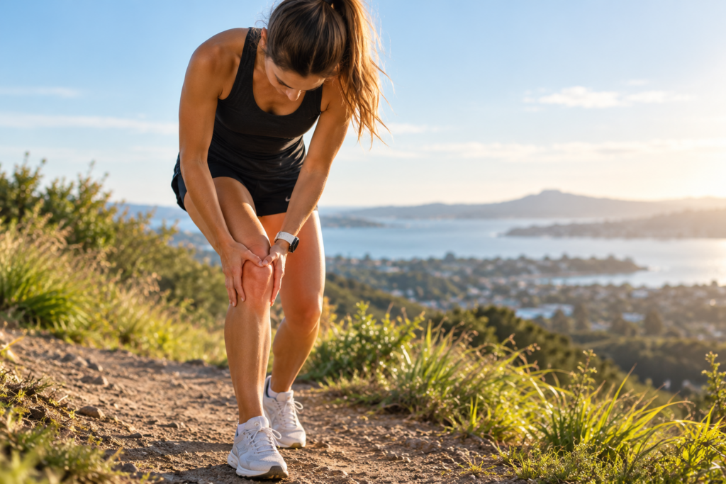
A deep, dull ache behind or around the kneecap (patella) is one of the most common complaints among trail and road runners. Known clinically as Patellofemoral Pain Syndrome (PFPS)—and casually as “Runner’s Knee”—this condition frequently flares up during hill sessions.
Whether pushing up a steep incline or controlling a fast descent, hill running imposes distinct biomechanical demands on the patellofemoral joint. Understanding why tracking issues occur during elevated runs is the key to resolving the pain for good.
The Biomechanics: Why Hills Exacerbate PFPS
The patella acts as a pulley system inside the trochlear groove of the femur. Under normal conditions, it glides smoothly as the knee flexes and extends. However, when biomechanical tracking is altered, excessive compressive force is directed onto the retropatellar cartilage and subchondral bone.
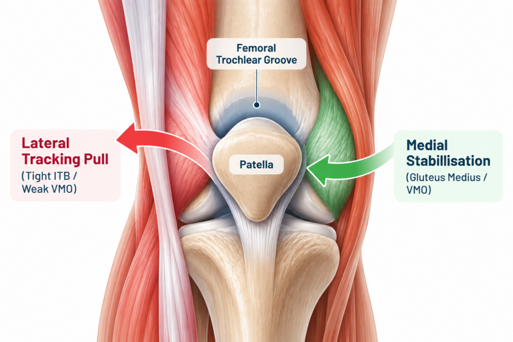
Hill running amplifies these compressive stress vectors in two distinct ways:
- Uphill Running (High Knee Flexion & Quad Demand): Propelling your bodyweight uphill requires greater knee flexion angles and maximal quadriceps contraction. As knee flexion increases beyond 30°, patellofemoral joint reaction forces (PFJRF) spike dramatically.
- Downhill Running (Eccentric Braking Forces): Downhill running relies heavily on eccentric quadriceps absorption. The muscle works while lengthening, driving high impact forces directly through the patellofemoral interface with every footstrike.
Root Causes of Mal-Tracking in Runners
PFPS is rarely an isolated knee problem; it is typically a proximal (hip) or distal (foot) mechanics issue manifesting at the knee joint.
1. Proximal Deficits (Hip & Gluteal Weakness)
Weakness in the gluteus medius and gluteus maximus allows hip adduction and internal rotation during single-leg stance. This dynamic collapse—often called dynamic knee valgus—causes the femur to rotate underneath the kneecap, effectively pulling the trochlear groove away from a centered patella.
2. Local Muscle Imbalances
An imbalance between the lateral structures (tight Iliotibial Band / Tensor Fasciae Latae) and the medial stabilizers (weak Vastus Medialis Obliquus / VMO) pulls the patella laterally against the outer wall of the femoral groove.
3. Distal Mechanics (Foot Pronation)
Excessive or prolonged foot pronation drives internal tibial rotation, which in turn alters knee kinematics during stance phase, putting added torque on the extensor mechanism.
Evidence-Based Rehabilitation Framework
Fixing patellofemoral pain requires a targeted load-management and progressive strengthening approach rather than passive rest alone.
Phase 1: Load Modification & Acute Management
- Cadence Adjustment: Increasing running cadence by 5–10% reduces peak knee flexion angles and decreases patellofemoral joint load per step.
- Temporary Slope Reduction: Avoid steep downhill running during acute flare-ups; transition to flat terrain or low-incline treadmills to manage acute stress.
- Targeted Taping: Patellar taping (McConnell technique) can offer short-term pain relief, allowing runners to engage in strength training comfortably.
Phase 2: Targeted Resistance Training
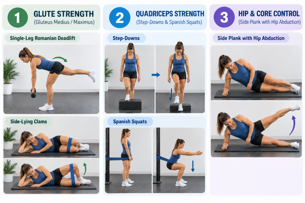
Strength exercise selection should focus heavily on hip abduction, external rotation, and closed-kinetic-chain quadriceps loading:
| Muscle Group | Primary Exercise | Focus / Target |
| Gluteus Medius / Maximus | Single-Leg Romanian Deadlifts & Side-Lying Clams | Prevents dynamic valgus and femoral internal rotation. |
| Quadriceps / VMO | Step-Downs & Spanish Squats | Heavy, slow resistance (HSR) training to build load tolerance. |
| Core & Trunk Stabilizers | Side Planks with Hip Abduction | Controls pelvic drop (Trendelenburg gait) during footstrike. |
When to Consult a Physiotherapist
If anterior knee pain persists despite load management, a clinical assessment can pinpoint exact structural or kinematic drivers. A physical examination evaluates:
- Running gait kinematics via video analysis.
- Quadriceps and gluteal force production profiles.
- Patellar mobility and soft-tissue restrictions.
References
- Crossley, K. M., et al. (2016). 2016 Consensus statement on exercise therapy and physical therapy interventions to treat patellofemoral pain. British Journal of Sports Medicine, 50(14), 839-843.
- Willy, R. W., et al. (2019). Patellofemoral Pain: Clinical Practice Guidelines Linked to the International Classification of Functioning, Disability and Health. Journal of Orthopaedic & Sports Physical Therapy, 49(9), CPG1-CPG95.
- Neal, B. S., et al. (2016). Six key features of patellofemoral pain syndrome: A systematic review. Gait & Posture, 45, 227-235.
- Barton, C. J., et al. (2015). The Patellofemoral Pain Clinical Framework: a consensus statement on biomechanical assessment and intervention. British Journal of Sports Medicine, 49(21), 1350-1357.